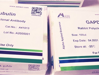

Bestrophin-1 rabbit pAb
Cat :A11212
-
Source
Rabbit
-
Applications
WB,IHC
-
Reactivity
Human,Rat,Mouse
-
Dilution
WB: 1:500 - 1:2000. IHC: 1:100 - 1:300. ELISA: 1:40000. Not yet tested in other applications.
-
Storage
-20°C/1 year
-
Specificity
The antibody detects endogenous Bestrophin-1 protein
-
Source/Purification
The antibody was affinity-purified from rabbit antiserum by affinity-chromatography using epitope-specific immunogen.
-
Immunogen
Synthetic Peptide of Bestrophin-1
-
Uniprot No
O76090
-
Alternative names
Bestrophin-1 (TU15B) (Vitelliform macular dystrophy protein 2)
-
Form
Liquid in PBS containing 50% glycerol, 0.5% BSA and 0.02% sodium azide.
-
Clonality
Polyclonal
-
Isotype
IgG
-
Background
bestrophin 1(BEST1) Homo sapiens This gene encodes a member of the bestrophin gene family. This small gene family is characterized by proteins with a highly conserved N-terminus with four to six transmembrane domains. Bestrophins may form chloride ion channels or may regulate voltage-gated L-type calcium-ion channels. Bestrophins are generally believed to form calcium-activated chloride-ion channels in epithelial cells but they have also been shown to be highly permeable to bicarbonate ion transport in retinal tissue. Mutations in this gene are responsible for juvenile-onset vitelliform macular dystrophy (VMD2), also known as Best macular dystrophy, in addition to adult-onset vitelliform macular dystrophy (AVMD) and other retinopathies. Alternative splicing results in multiple variants encoding distinct isoforms.[provided by RefSeq, Nov 2008],
-
Other
BEST1, Bestrophin-1 (TU15B) (Vitelliform macular dystrophy protein 2)
-
Concentration
1 mg/ml
| Product | Reactivity | Applications | Conjugation | Catalog | Images |
|---|
-
 400-836-3211
-
 support@aabsci.com
-
β-actin rabbit pAb ...... >
-
β-actin rabbit pAb(A284) ...... >
-
Plant-actin rabbit pAb ...... >
-
β-tubulin mouse mAb(M7) ...... >
-
GAPDH mouse mAb(2B8) ...... >
-
GAPDH mouse mAb(PT0325) ...... >
-
Histone H3 rabbit pAb ...... >
-
Histone H3 rabbit pAb ...... >
-
COX IV mouse mAb(6C8) ...... >
-
GFP-Tag mouse mAb(1G6) ...... >
-
HA-Tag mouse mAb(1B10) ...... >
-
mCherry-Tag mouse mAb(6B3) ...... >


 400-836-3211
